-

★:There is a signboard to guide you to our clinic.
-
〒121-0061
Yokoyama Eye Building 1F・2F, 1-7-19 Hanabata, Adachi-ku, TokyoTEL
03-3885-0412
Closed: Wednesdays, national holidays, summer holidays, year-end and New Year holidays, and others.
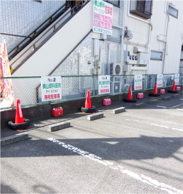
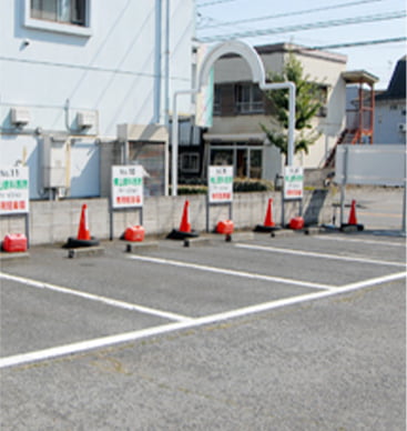
- parking lot:20 units
- parking area for bicycles:20 units available
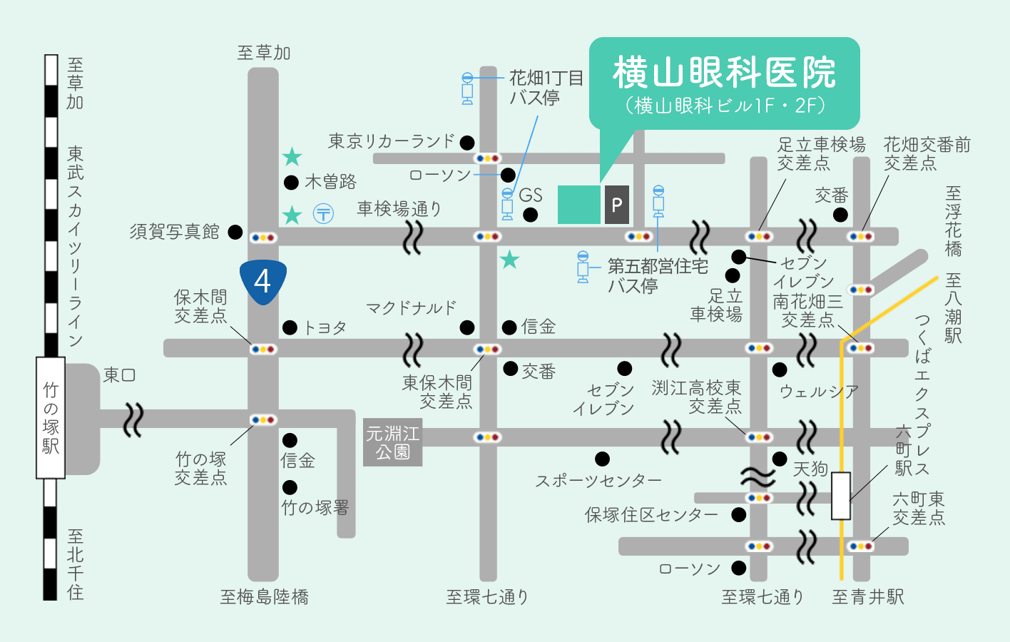
For those coming by public transportation
- Take the Tobu Bus from the east exit of Takenotsuka Station.
-
- Aya24: Take the bus via Rokucho Station and Shakenjo (bound for Ayase Station) and get off at the 5th Toei Jutaku bus stop.
- Tobu Bus from Mu-machi Station
-
- Aya24, via Rokucho Station and Shakenoba (bound for Takenotsuka Station East Exit), get off at Fifth Toei Jutaku bus stop.
- Tobu Bus from Ayase Station
-
- Aya24, via Rokucho Station and Shakenoba (bound for Takenotsuka Station East Exit), get off at Fifth Toei Jutaku bus stop.
- Aya 40 (for Hanabatake Danchi), Hanabatake 1-chome stop, 2 minutes walk
Facilities
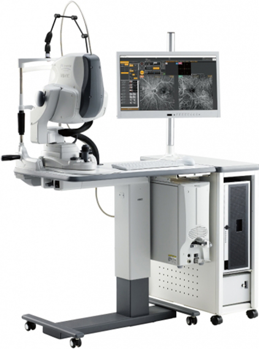
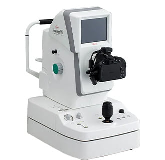
I introduce cofocus scanning type diode laser eye examination "Mirante" two
I introduce range, high resolution that are wider than conventional fundus camera, diode laser eye examination apparatus "Mirante" which I can photograph speedily two at our hospital.
Because this apparatus does not have to open the pupil in the case of inspection, patient that you came by car can undergo an examination of the eyeground.
-
Characteristic of Mirante.1
Eyeground shooting is possible without even a person coming by car dilating.
-
Characteristic of Mirante.2
Taking picture of photograph which is higher-resolution than a conventional apparatus is possible.
-
Characteristic of Mirante.3
I can photograph retinal blood vessels and I in this way discover changes such as the abnormal blood vessel or falling off early and grasp it before diabetes retinopathies are aggravated and tie it to appropriate treatment.
-
- optical coherence tomography(OCT)
- It can observe the shape and lesions of the retina and measure the thickness of the nerves.
It is also possible to take images of blood vessels without the use of contrast media.
-
- non-mydriatic fundus camera
- This instrument uses infrared light to observe and photograph the fundus of the eye.
-
- Specular Microscope
- This device is used to photograph and analyze corneal endothelial cells, which help keep the cornea transparent.
-
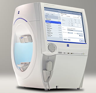
- Optical biometric device
- 白This instrument can measure the ocular axis length (length of the eye), corneal roundness, anterior chamber depth, and lens thickness, which are necessary for cataract surgery, all at once without touching the eye.
-
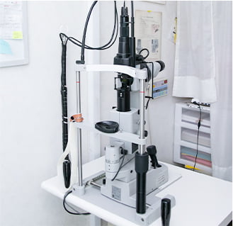
- laser
- Laser therapy is now a common treatment method in the field of ophthalmology. The equipment used and the irradiation method vary depending on the disease.
-
- YAG Laser Surgery System
- This YAG laser system is used for surgery to drill holes in the iris for the treatment of posterior cataracts and glaucoma. The treatment can be performed safely and efficiently.
-
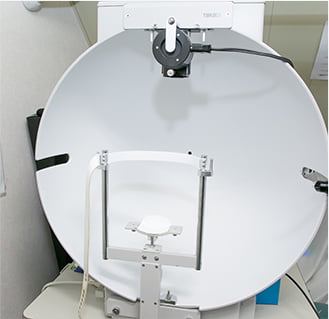
- Large amblyopia mirror
- It is used to examine and treat strabismus and amblyopia.
Measures the position of the eyes and whether the patient is able to see in three dimensions with both eyes.
-
- Humphrey Visual Field Meter
- This device detects minute visual field abnormalities in the central visual field, including glaucoma.
<2 units>
-
- Goldmann Visual Field Analyzer
- This perimeter is effective in detecting a variety of visual field abnormalities. Flicker stimulation makes it possible to detect early damage to the third neuron, such as optic neuropathy.
-
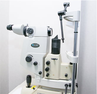
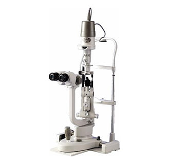
- small-gap light microscope
- By adjusting the type and angle of the light, the surface of the eye, conjunctiva, cornea, and lens are examined.
-
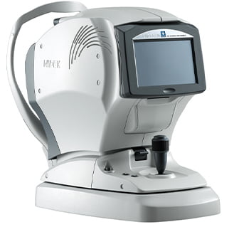
- Auto-reflex kerato/tono/pachymeter
- A variety of functions such as refraction, corneal radius of curvature measurement, intraocular pressure measurement, corneal thickness measurement, and adjustment force measurement are now possible with a single device.
<3 units>
-
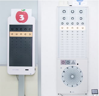
- space saving chart
- This vision chart allows you to test the equivalent of 5 meters at a distance of 1 meter.
It also has the feature that you can perform the test without letting others know your eyesight.
-
- eyesight test chart
- An eye exam is one of the basic tests performed in an eye clinic. You will be asked to look at the Landolt ring one eye at a time, and your visual acuity will be measured according to the size of the ring you can see.
-
- Binocular Vision Testers
- This is a device that relaxes children's eyes by having them look at the scenery displayed inside the Wack, which helps their eyes become accustomed to looking at nearby objects when operating a smartphone or reading.
-
- Electric Deep Vision Meter
- When obtaining or renewing a large vehicle or a second class driver's license, you will need to pass a test to determine your perspective. If you would like to practice using the depth meter, please ask our staff for more information.
<Depth inspection and training such as large scale training is possible.>
-
- Handy fundus camera
- Take a hand-held fundus photograph.
The examination can be performed in a wheelchair.
-
- Handy eye pressure system
- Hand-held IOP measurement.
The examination can be performed in a wheelchair.
-
- Handy Auto Reflex Kerat
- Hand-held refractive index measurement and corneal radius of freedom measurement.
The examination can be performed in a wheelchair.
-
- center flicker
- The patient is asked to look at a discontinuous light that repeatedly blinks and measures whether there is any abnormality in the optic nerve function.